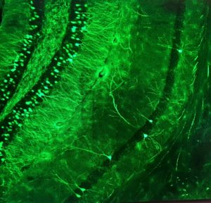
 Optical recording methods have been refined to the point where they are very useful in measuring neuronal activity. Optical imaging enables the assessment of more neurons than traditional electrophysiological methods and provides spatial/anatomical information that cannot be deciphered from an electrode. Calcium indicators have been employed in the hippocampus of awake and behaving animals and have answered questions about the relative stability and plasticity of hippocampal neuronal ensembles. However, calcium signals are slow and often cannot resolve individual action potentials. GEVIs overcome the temporal problem and are now very bright, but a lack of compartmentalization severely compromises the signal to noise ratio in dense mammalian circuits. We are working to localize GEVIs to the neuronal soma for the imaging of action potentials. The most promising constructs will be incorporated into viral vectors to express the soma-localized GEVIs in the hippocampus in vivo to study network operations in hippocampal slices and, eventually, to observe real-time network activity in mice as they perform spatial learning and memory tasks.
Neuronal Localization of Genetically-Encoded Voltage Indicators & Actuators
- Home
- /
- Neuronal Localization of Genetically-Encoded Voltage Indicators & Actuators